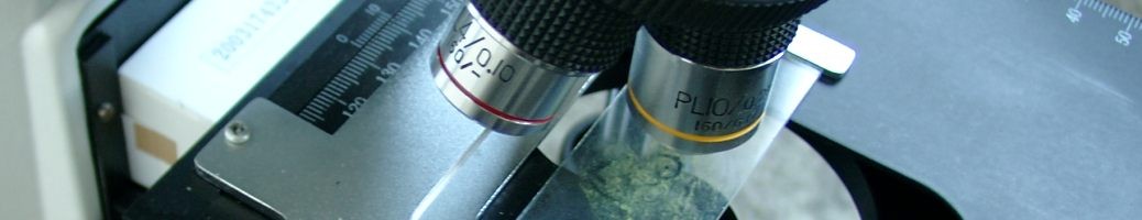
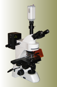
Figure 1: Microscope Micom E for algal growth inhibition test of toxicity evaluation
MICOM E enhances the Diram s.r.o instrument range to allow evaluation of algal growth inhibition according to the ISO8692 test of toxicity using green algae Desmodesmus subspicatus. Deploying image analysis on an eluate sample on a Bürker counting chamber, the MICOM E allows fast, accurate assessment.
The employed analytical method relies on observing the light re-emitted (λ=682nm) by the chlorophyll of living algae cells after excitation from a narrow-band blue light (λ=450nm).
The disadvantage of this method is the potential bleaching of the chlorophyll and the resultant decrease in fluorescence intensity after several tenths of a second. To avoid the degradation, MICOM E moves the stage and captures images in a sufficiently short time frame. Then software analyses the number of cells and their dimensions. Eventual conglomerates of cells can then be separated to obtain the number of living cells per sample volume.
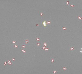
Figure 2: Image analysis of fluorescence of living cells
The MICOM E package combines a fluorescence microscope with trinocular tubus and motorised stage, a CCD camera, and the Lambdasoft software to control stage movement and image capture. Lambdasoft also allows control of camera parameters such as brightness, contrast, and exposure; and various advanced image analysis factors (thresholding, filtration, size determination method…). It includes user database for saving and export of images.